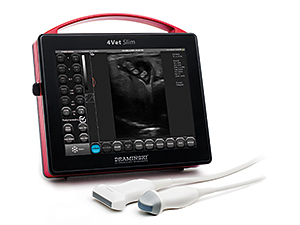
ULTRASOUND SCANNER 4Vet Slim

ULTRASOUND SCANNER 4Vet Slim
Ultrasound scanner with a high quality imaging for the diagnosis of small animals and horses. Perfect for veterinary mixed practises.
This ultrasound scanner combines elegance, simplicity and functionality.
Why is it worth having the DRAMINSKI 4Vet Slim?
Technologies of the future
- ULTRASOUND IMAGING IN ANY LIGHTING CONDITIONS
- INDEPENDENCE
- MODERN DESIGN
- SIMPLICITY
- FUNCTIONALITY
- SAFETY
Obtain a clear image no matter where you are, thanks to a high contrast, bright screen.
Perform examination even with limited access to electricity. A more powerful battery pack enables 4 hours of work on a single charge.
Combination of aluminium and glass, manual finishing of every detail enchants with the shape and elegance. All that is to make your customers feel exceptional.
All the necessary functions are at hand, a sensitive touch screen makes setting of a proper image faster.
After 25 seconds Slim is ready for operation. A wide choice of the probes enables examination of different patients. Echo enhancement of the needle facilitates injections, and the colour Doppler helps differentiate the blood vessels from other structures. Do you need anything more for effective testing?
Two-year warranty, for both the device and the probes, gives a sense of comfort of work. We know how important it is because we have been working together with veterinarians for many years.
Technical Data
| External dimensions | 26 cm x 25 cm x 5.5 cm |
| Weigh | 2800 g |
| Battery weight | 780 g |
| Type of imaging |
B Mode B+B Mode 4B Mode M Mode B+M Mode Color Doppler Power Doppler (PDI) Pulse Wave Doppler (PWD) |
| Frequency | 2-14 MHz (depends on the type of a probe) |
| Dynamic focusing | yes |
| Image managing |
– Freeze – Zoom 60 – 300% interval every 20% – Full screen – Saving images and cine loop (256 frames) |
| Presety | improvement of the needle visibility in images from the linear probe; abdominal cavity cat, abdominal cavity middle-size dog, abdominal cavity big dog, mare pregnancy, mare ovary, mare uterus, horse tendons |
| Grey scale | 256 degrees |
| Post-processing regulation | on/off |
| User panel | – menu language version: Polish, English, Spanish, French, German, Russian, Croatian, Arabic, Korean – optimization of the image setting parameters |
| Measuring options | – standard package: ruler, grid, distance, volume, surface area, ellipsis, narrowing; – OB./GYN package: age tables for different species of animals |
| System | integrated with PC |
| Monitor | LCD LED display, diagonal 10.4” |
| Control of functions | capacitive touch screen |
| Memory of images and cine loop |
100 GB saving of the images and cine loop with description, patient data and date |
| Data transfer standard | DICOM 3.0 |
| Data transmission to the external carrier | via USB |
| Number of probe ports | one port, automatic probe recognition |
| External ports | 2 x USB 3.0, 1 x LAN, 1 x Display Port |
| Source of power supply | 1. Power supply; input: 100-240V AC, 50-60Hz, max 1.5A output: 18V DC / 3.34A 2. Li-Ion battery pack, 14.4V, 10Ah |
| Time of continuous work at battery supply |
about 4 hours |
| Time of battery charging | about 3 hours (battery charger type: 3240 LI) |
| Battery discharge indicator | graphic battery discharge indicator |
| Work readiness time | about 25 s |
| Enclosure | metal: duraluminium |
| Operating temperature | -15°C to +45°C |
| Storage temperature | 0°C to +45°C |
| Current consumption | about 2.2 A |
| Optional accessories | Wive-wheel trolley (two of them can be blocked) horizontally and vertically adjustable, the scanner can be bent horizontally. It has a basket and holders for the probes. |
| Usage | ultrasound diagnostics of animals |